Program
Follow this link for the detailed program.
See the Program Booklet for general information and detailed program.
For the book of abstracts, please follow this link.
For the Photo Contest Booklet, please follow this link.
Plenary Speakers
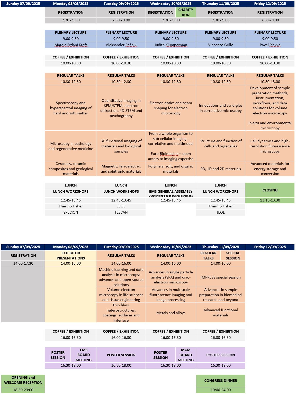
Vincenzo Grillo
Instituto Nanoscienze of the National Research Council, Modena, Italy
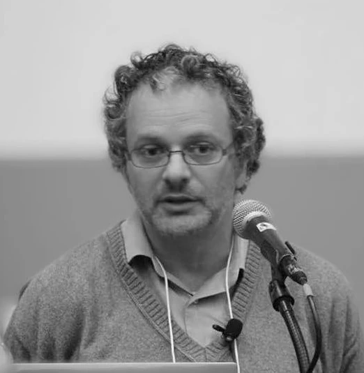
Mateja Erdani Kreft
Institute of Cell Biology, University of Ljubljana, Slovenia
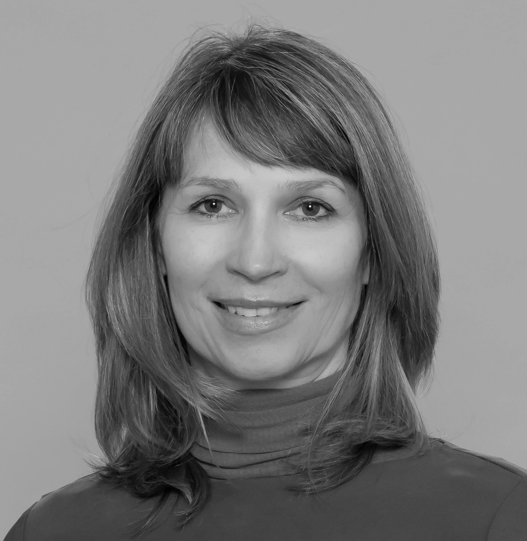
Aleksander Rečnik
Jožef Stefan Intitute, Ljubljana, Slovenia
Pavel Plevka
CEITEC – Central European Institute of Technology, Brno, Czech Republic

Judith Klumperman
Center for Molecular Medicine Section Cell Biology, University Medical Center Utrecht, the Netherlands
Instrumentation and Methods
Spectroscopy and hyperspectral imaging of hard and soft matter
This symposium explores the latest developments and advanced applications of spectroscopy and hyperspectral imaging for the analysis of hard and soft materials. Aiming to improve the characterisation of surfaces and interfaces, participants will discuss advances in spectroscopic techniques and hyperspectral imaging for the determination of chemical composition, structure and functional properties. Innovative approaches for multi-scale and multidimensional analysis will be addressed, with a focus on the fields of advanced materials, and nanoscience.
Chairs
- Michael Stöger-Pollach, Vienna University of Technology, Vienna, Austria
- Giuseppe Nicotra, CNR Institute for Microelectronics and Microsystems, Catania, Italy
Invited speakers
- Quentin Ramasse, Superstem Lab Daresbury UK
- Odile Stéphan, University of Paris Saclay, Gif-sur-Yvette, France
Innovations and synergies in correlative microscopy
The session “Innovations and Synergies in Correlative Microscopy” will highlight cutting-edge advancements in microscopy techniques, featuring two distinguished speakers. Prof. Alexandre A. Mironov from IFOM (Istituto Fondazione di Oncologia Molecolare) will discuss his pioneering work in ultrastructural analysis using correlative light and electron microscopy (CLEM) to study cell biology in health and disease. His research focuses on the role of cellular organelles and processes in physiological and pathological states, employing advanced EM methods like CLEM and three-dimensional EM to uncover cellular alterations invisible under light microscopy. Dr. Roman Polishchuk, Laboratory Head at TIGEM (Telethon Institute of Genetics and Medicine), will share his contributions to characterizing intracellular membrane trafficking and cell polarity in genetic disorders and other pathologies. He has been instrumental in developing CLEM technology, integrating live cell imaging with EM to advance our understanding of cellular processes. We look forward to excellent scientific contributions in the field of advanced correlative microscopy and to an exciting discussion.
Chairs
- Philip Steiner, Johannes Kepler University Linz, Austria
- Mateja Erdani Kreft, Institute of Cell Biology, University of Ljubljana, Slovenia
Invited speakers
- Alexandre A. Mironov, IFOM ETS, Milan, Italy
- Roman Polishchuk, Telethon Institute of Genetics and Medicine (TIGEM), Pozzuoli, Italy
Quantitative imaging in SEM/STEM, electron diffraction, 4D-STEM and ptychography
The session allows for precise measurements of structural parameters, strain fields, and electromagnetic properties, and enable the detailed structural and compositional analysis, guiding the design of new materials. The integration of machine learning and advanced computational algorithms enrich these methods further and lead to more accurate and efficient data interpretation. The session aims to highlight the latest developments in all aspects of computational and experimental microscopy.
Chairs
- Gerald Kothleitner, FELMI, Graz University of Technology, Graz, Austria
- Giovanni Bertoni, Istituto Nanoscienze, CNR, Modena, Italy
Invited speakers
- Lothar Houben, Weizmann Institute of Science, Israel
- Benedikt Haas, HU Berlin, Germany
Advances in single particle analysis (SPA) and Cryo-Electron Microscopy
Recent advancements in Single Particle Analysis (SPA) and Cryo-Electron Microscopy (Cryo-EM) have revolutionized structural biology, enabling near-atomic and sub-angstrom resolution of biological macromolecules.
The integration of direct electron detectors, AI-driven image processing, and energy filter technology has significantly enhanced data quality and resolution. Cryo-Electron Tomography (Cryo-ET) enables in situ structural analysis, extending Cryo-EM beyond isolated proteins to complex cellular environments. Advances in automated sample preparation and high-throughput data collection are further democratizing the technique, reducing technical barriers and making it more accessible to a broader research community. As computational and instrumental innovations continue, Cryo-EM is set to further transform molecular and cellular biology, bridging the gap between structure and function. This session will highlight cutting-edge Cryo-EM applications across various scientific fields, showcasing its impact on molecular and cellular research. Join us for an in-depth discussion on the latest breakthroughs and future directions in this rapidly evolving field.
Chairs
- Kamila Hrubanová, Institute of Scientific Instruments of the Czech Academy of Sciences, Brno, Czech Republic
- Gašper Šolinc, National Institute of Chemistry, Ljubljana, Slovenia
Invited speakers
- Marjetka Podobnik, National Institute of Chemistry, Ljubljana, Slovenia
- Pavel Křepelka, CEITEC, Brno, Czech Republic
Development of sample preparation methods, instrumentation, workflows, and data solutions for volume electron microscopy
Volume electron microscopy enables the generation of high-resolution 3D models of objects at the nanoscale, providing valuable insights into underlying processes. The successful application of these methods requires a thorough understanding and precise execution of all essential steps, including specimen preparation, instrumentation, data acquisition, processing, and data storage. In this session, we welcome contributions that present innovative strategies for optimizing any of these steps to enhance the quality and reliability of results.
Chairs
- Jana Nebesářová, Biology Centre of the Czech Academy of Sciences, České Budějovice, Czech Republic
- Barbara Šetina Batič, Institute of Metals and Technology, Ljubljana, Slovenia
Invited speakers
- Miroslav Šlouf, Institute of Macromolecular Chemistry, Prague, Czech Republic
- Stefan Zaefferer, Max Planck Institute for Sustainable Materials (MPIE), Duesseldorf, Germany
Electron optics and beam shaping for electron microscopy
The incredible success of electron microscopy in recent years builds, aside from improvements in detector technology and data processing, on innovation in electron optics. We now have much better spatial and energy resolution, more detailed chemical and analytical information, spectroscopy of excitations, imaging of dose sensitive material than even 10 years ago, permitting us to explore materials phenomena that were previously inaccessible or accessible less directly by other probes.
Beyond this, the emerging capability of shaping electron beams with very many degrees of freedom takes us beyond the paradigm of standard electron optics. Inspired by light optics in the capability to arbitrarily modify a coherent wavefront, beam shaping allows us to obtain functional probes and novel detection methods.
This session is therefore dedicated to the innovation in both conventional electron optics and electron beam shaping (in both space and time?). Special attention will be given to techniques that push the boundary of microscopy in the direction of quantum metrology.
Chairs
- Christoph Koch, Humboldt University of Berlin, Berlin, Gremany
- Vincenzo Grillo, Instituto Nanoscienze of the National Research Council, Modena, Italy
Invited speakers
- Thomas Juffman, Vienna University, Vienna, Austria
- Axel Lubk, Leibniz Institute for Solid State and Materials Research (IFW), Dresden, Germany
In-situ and environmental microscopy
Structural and functional materials often operate under dynamic conditions, such as elevated temperatures, electrical and magnetic fields, mechanical loads, and under various aqueous, gaseous and electrochemical environments. Emerging in-situ and environmental microscopy techniques are transforming multidisciplinary research by providing unprecedented insights into material behaviour under realistic in-situ and operando conditions. These advancements are revolutionising experimental platforms across physics, chemistry, materials science, and beyond. Innovations such as advanced aberration correctors, functional TEM/SEM holders, and ultra-sensitive detectors enable multi-scale, temporal, and high spatial resolution analyses, unlocking previously unattainable understanding of structural, optical, mechanical, and chemical processes in materials.
Contributions on all TEM/SEM methods for investigating dynamic processes are welcomed. Relevant topics include, but are not limited to, structural and functional materials, such as energy materials, catalytic processes and chemical reactions, low-dimensional nanomaterials, and geological materials and processes.
Chairs
- Sašo Šturm, Jožef Stefan Institute, Ljubljana, Slovenia
- Zaoli Zhang, Erich Schmid Institute of Materials Science, Austrian Academy of Sciences, Leoben, Austria
Invited speakers
- Jinming Guo, School of materials science and engineering, Hubei University, Wuhan, China
Machine learning and data analysis in microscopy: Advances and open-source solutions
Open-source software has emerged as a vital catalyst in the advancement of electron microscopy. By empowering researchers to collaboratively develop and refine tools with the advanced features necessary for cutting-edge investigations, it accelerates scientific discovery while ensuring transparency and reproducibility. Moreover, the integration of machine learning and artificial intelligence into data acquisition, analysis, simulation, and interpretation is revolutionising the field. These approaches are transforming the way we process and understand complex, multi-dimensional datasets generated by modern microscopes.
This session will explore the latest advances in open-source solutions and applications of machine learning to electron microscopy.
Chairs
- Francisco de la Peña, Lille University, Lille, France
Invited Speakers
- Diana Propst, Faculty of Physics, University of Vienna, Vienna, Austria
Life Sciences
Advances in multiscale fluorescence imaging and image processing
In this session fundemental challenges in multiscale fluorescence imaging and image processing along with successful exemples will be discussed to highlight the effectiveness of this approach. Fluorescence microscopy is a very effective tool to study biological structure and function of the cell. Multiscale fluorescence imaging is the capability to traverse multiple spatial or temporal scales when performing a microscopy experiment. In order to be able to examine cellular behaviour in various spatial dimensions and over time, different imaging technologies are incorporated to the investigations. The integration of the information gathered from the individual cellular level up to the level of the entire organism will allow to identify links between cellular mechanisms and the function of organs to permit inspection of dynamics over diferent timescales. New challenges are needed in order to integrate data sets from different imaging techniques to recognise complex patterns in cell behaviour on a holistic level.
Chairs
- Serap Arbak, Department of Histology and Embryology, Acibadem University, Istanbul, Turkey
Invited speakers
- Joerg Enderlein, Institute of Physics of the Georg August University Göttingen, Germany
Volume electron microscopy in life sciences and tissue engineering
This session delves into two interconnected themes: the use of volume electron microscopy (vEM) in life sciences and advancements in tissue engineering. For life sciences, the session highlights vEM’s role in unravelling complex 3D structures of tissues and ultrastructures of cells and subcellular compartments, bridging the gap between structural resolution and functional insight. Key discussions include innovations in imaging workflows, integration with other modalities, and analysis pipelines designed for large-scale datasets. In the realm of tissue engineering, the focus shifts to exploring cutting-edge strategies for designing, visualizing, and analyzing engineered scaffolds and cell-material interactions. While these two fields often operate independently, the session emphasizes the active involvement of both, encouraging direct collaboration. By bringing together researchers in the fields of vEM and tissue engineering, the session aims to foster a dynamic exchange of ideas, techniques, and solutions that have the potential to drive new breakthroughs at the intersection of these disciplines.
Chairs
- Jiří Týč, Biology Centre of the Czech Academy of Sciences, České Budějovice, Czech Republic
- Halime Kenar, Department of Biomedical Engineering, Acibadem University, Istanbul, Turkey
Invited speakers
- Jemima Burden, University College London, UK
- Duarte Barral, NOVA Medical School, Lisbon, Portugal
Structure and function of cells and organelles
This session aims to provide a comprehensive platform for sharing discoveries that enhance our understanding of cellular structure and function. We invite talks that apply various microscopic tools in any model organism (cell lines, invertebrates, vertebrates and plants) and present results that help to understand the architecture and dynamic functions of cells and their organelles. Studies on membrane trafficking and the emerging field of intra- and extracellular vesicles, including the biogenesis and functional roles of vesicles in intra- and intercellular communication, are requested. Autophagy and related processes hold special interest. Topics involving the combination of state-of-the-art microscopy technologies are warmly welcomed. Works that uncover novel regulatory pathways with structural consequences, signalling/secretion mechanisms, and their implications in health and disease is encouraged.
Chairs
- Tamás Visnovitz, Semmelweis University, Budapest, Hungary
- Petra Peharec Štefanić, Division of Molecular Biology, Department of Biology, Faculty of Science, University of Zagreb, Croatia
Invited speakers
- Péter Lőrincz, ELTE Eötvös Loránd University, Budapest, Hungary
- Tea Mišić Radić, Ruđer Bošković Institute, Zagreb, Croatia
Microscopy in pathology and regenerative medicine
This session will deal with topics ranging from pathology research and diagnostics to regenerative medicine, with the common background of microscopy. The usefulness of various studies based on light microscopy, transmission/scanning electron microscopy, and fluorescent/confocal techniques will be highlighted and represent the core of this panel. We anticipate a significant contribution from pathologists presenting a broad panel of antibodies used in diagnostics. Also, scientists engaged in research on regenerative medicine are cordially invited to participate and present their results, sharing and introducing new directions, trends, and achievements in up-to-date medicine, dentistry, and veterinary medicine.
Chairs
- Elena Bianca Donetti, University of Milan, Milan, Italy
- Nela Puškaš, Institute of Histology and Embryology, Faculty of Medicine, University of Belgrade, Belgrade, Serbia
Invited speakers
- Ida Perrotta, University of Calabria, Italy
- Ivan Čapo, University of Novi Sad, Faculty of Medicine, Novi Sad, Serbia
Cell dynamics and high-resolution fluorescence microscopy
Fluorescence microscopy remains an indispensable tool for investigating subcellular structures and dynamic processes in live samples. By enabling real-time, non-invasive imaging, this technique provides insights into cellular mechanisms under physiological and pathological conditions. Recent advancements in far-field fluorescence microscopy, particularly the emergence of high-speed methods, such as the spinning disk microscopy, and super-resolution methods such as STED, SIM, and STORM, have redefined the limits of optical microscopy. These techniques have significantly improved temporal resolution and achieve spatial resolutions down to a few tens of nanometers, far exceeding the classical diffraction limit (~200 nm laterally and ~600 nm axially), thereby bridging the resolution gap between light and electron microscopy. Enhanced spatial resolution of super-resolution techniques has significantly advanced our understanding of cellular and tissue architecture. Furthermore, the ability to track live processes at these scales and improved speed has provided invaluable insights into dynamic cellular events. The session will showcase these advancements, highlighting their impact on the fields of cell biology and physiology.
Chairs
- Jernej Jorgačevski, Institute of Pathophysiology, Faculty of Medicine, University of Ljubljana, Ljubljana, Slovenia
- Maja Herak Bosnar, Division of Molecular Medicine, Ruđer Bošković Institute, Zagreb, Croatia
Invited speakers
- Marko Kreft, Biotechnical Faculty, University of Ljubljana, Slovenia
- Vedrana Filić Mileta, Ruđer Bošković Institute, Zagreb, Croatia
From a whole organism to sub-cellular imaging: correlative microscopy and multimodal imaging
Correlative microscopy and multimodal imaging integrate various modalities based on different energy sources, such as light, electrons and X-rays, to study the same object. These techniques produce images across a wide range of scales—from molecular details to entire organisms. While each imaging tool has its advantages and limitations, combining their outputs allows researchers generally to overlay structural, functional, and compositional data. This integration places findings within a precise anatomical/structural context, enabling a deeper understanding of biological processes and their functions. Join us to explore innovative workflows, strategies and solutions in correlative and multimodal imaging. Researchers from all disciplines are invited to participate!
Chairs
- Polona Mrak, Biotechnical Faculty, University of Ljubljana, Ljubljana, Slovenia
- Marie Vancová, Biology Centre of the Czech Academy of Sciences, České Budějovice, Czech Republic
Invited speakers
- Jernej Jorgačevski, Institute of Pathophysiology, Faculty of Medicine, University of Ljubljana, Ljubljana, Slovenia
- Jiří Týč, Biology Centre of the Czech Academy of Sciences, České Budějovice
Advances in sample preparation in biomedical research and beyond
Microscopy is currently undergoing a revolution driven by advanced techniques such as 3D/volume EM, cryo-EM, live cell imaging, CLEM, super-resolution and others. However, time spent behind the microscope is just the tip of the iceberg in the broader workflow of sample analysis. Most scientists agree that sample preparation is crucial in determining the quality and relevance of the data. Sample preparation is often the most experience-demanding, time-consuming and complex part of the process, yet is sometimes somewhat neglected and the effort involved is often underestimated.
This session is dedicated to highlighting recent advances in sample preparation for biomedical research and life sciences in general. Researchers are encouraged to share their work, focusing on novel approaches in the field, best practises and lessons learnt, but also on challenges and real-world obstacles encountered at any stage of sample preparation. Topics of interest include, but are not limited to, cryo- and chemical fixation, freeze-substitution protocols, embedding media selection for immunolabelling or ultrastructural research, correlative microscopy, organoid imaging, live cell imaging, visualization of extracellular vesicles and similar.
Chairs
- Samo Hudoklin, Institute of Cell Biology, University of Ljubljana, Ljubljana, Slovenia
- Milica Markelić, Department of Cell and Tissue Biology, University of Belgrade – Faculty of Biology, Serbia
Invited speakers
- Marie Vancová, Biology Centre of the Czech Academy of Sciences, České Budějovice, Czech Republic
- Roland Fleck, King’s College London, UK
3D functional imaging of materials and biological samples
While we live in a three-dimensional world, traditional imaging techniques, including microscopy, have often been limited to two-dimensional views. Recent advances in microscopy, labeling techniques, and computational analysis have bridged this gap, enabling detailed 3D visualization of objects across a range of spatial and temporal scales.
This session will highlight recent progress in 3D imaging and analysis, focusing on morphological, temporal, and functional studies. Topics will include techniques such as X-ray microtomography, laser-scanning microscopy, super-resolution fluorescence microscopy, volume electron microscopy, and functional imaging methods like PET and MRI. Explore how these tools are shaping research and diagnostics today.
Although the session positioned within Life Science microscopy chapter, it will cover spatial imaging and analysis of organisms and materials.
Chairs
- Rok Kostanjšek, Biotechnical Faculty, University of Ljubljana, Ljubljana, Slovenia
- Gergely Szalay, Institute of Experimental Medicine, HUN-REN, Budapest, Hungary
Invited speakers
- Lucia Mancini, Slovenian National Building and Civil Engineering Institute, Ljubljana, Slovenia
- Kevin Gonzalez, Columbia University, New York City, USA
Material Sciences
Ceramics, ceramic composites and geological materials
All-scale microstructural characterization is essential for understanding the complex relationship between microstructure and properties in both synthesized and natural materials. The application of microscopy techniques drives innovation in ceramic materials and aids in interpreting the geophysical and geochemical history of minerals and rocks. In this session, we welcome contributions, where conventional and latest advanced microscopy methods including 2D and/or 3D imaging (SEM, TEM, STEM…), diffraction (EBSD, 4D STEM,…), spectroscopy (EDX, EELS, CL, Raman…) and FIB techniques, are applied at either normal or in-situ conditions to solving challenges in synthesized powders, polycrystalline ceramics and geological materials. Examples include, but are not limited to, characterization of microstructuralfeatures (distribution of grain sizes and grain orientation, analysis of phase composition, distribution of phases, the presence of inclusions, grain boundaries, planar defects like dislocations, twins, antiphase boundaries and others, the presence of oriented intergrowths and similar exciting micro- and nanoscale phenomena) to understand and further develop the properties of advanced and traditional ceramics and to understand mineral formation, diagenesis, and deformation in geological materials. This session invites contributions from academia, industry, and research institutions, fostering interdisciplinary discussions to address challenges and future directions in the field.
Chairs
- Servet Turan, Eskisehir Technical University, Eskisehir, Turkey
- Nina Daneu, Jožef Stefan Institute, Ljubljana, Slovenia
Invited speakers
- José Alberto Padron Navarta, Andalusian Institute of Earth Sciences, Granada, Spain
- Rik Drummond-Brydson, University of Leeds, Leeds, UK
Polymers, soft, and organic materials
The session invites contributions on both fundamental and applied aspects of polymers, biomaterials and other soft materials including polymer blends, composites, self-assembled particles in solutions, foams, hydrogels, liquid crystals, MOFs and COFs. The materials are expected to be investigated by state-of-the art microscopy techniques (namely light, electron, X-ray and scanning probe microscopy), but other characterization methods such as (micro/nano)spectroscopy and (micro/nano)mechanical characterizations are welcome as well.
Chairs
- Miroslav Šlouf, Institute of Macromolecular Chemistry, Prague, Czech Republic
- Rik Drummond-Brydson, University of Leeds, Leeds, UK
Invited speakers
- María de la Mata, University of Cádiz, Spain
- Daniel Horak, Czech Academy of Sciences, Prague, Czech Republic
Metals and alloys
This session is focused on microscopic studies of metals and their alloys, as well as intermetallics, metal matrix composites, and other metal-based materials.
The unique and novel advanced engineering metallic materials are attracting ever increasing attention and development for their exceptional combination of properties such as ultra high strength combined with lightweight, improved fatigue, corrosion resistance, biocompatibility and catalytic properties. To achieve this, the most important step is to determine microstructure – properties relationship, by studying the data obtained by microscopic examinations (LM, TEM, SEM, SPM, EDS, EBDS, diffraction patterns etc.) and their careful interpretation. Additionally, in the session, special cases of metal sample preparation might also be presented
Chairs
- Dragan Rajnović, Faculty of Technical Sciences, University of Novi Sad, Novi Sad, Serbia
- Matjaž Godec, Institute of Metals and Technology, Ljubljana, Slovenia
Invited speakers
- Alena Michalcova, Department of Metals and Corrosion Engineering, Prague
- Črtomir Donik, Institute of Metals and Technology, Ljubljana, Slovenia
0D, 1D and 2D materials
As nanomaterials play a central role in current and future technologies, advanced electron microscopy techniques have become essential for characterizing their structure, chemistry, and local properties. These materials include: nanoparticles, quantum dots, clusters, single atoms (all of them categorized as 0D), one-dimensional structures (such as nanowires, nanotubes, and nanorods), two-dimensional layered materials (graphene, TMDS, MXenes, etc.), and heterostructures. Electron microscopy is crucial for addressing emerging challenges in nanomaterials, like improving synthesis, controlling the size and morphology of nanoparticles, understanding the growth mechanisms and defects in 1D nanostructures, and examining the interfaces, strain, and electronic properties of 2D materials. This symposium will explore how advanced electron microscopy is being used to tackle these critical issues in nanomaterial characterization.
Chairs
- Mariana Klementová, Institute of Physics of the Czech Academy of Sciences, Prague
- Francisco Ruiz Zepeda, National Institute of Chemistry, Ljubljana, Slovenia
Invited speakers
- Paulo Ferreira, International Iberian Nanotechnology Laboratory, Braga, Portugal
- Sarah Haigh, The University of Manchester, Manchester, UK
Magnetic, ferroelectric, and spintronic materials
This section highlights advances in the study of magnetic, ferroelectric, and spintronic materials, emphasizing the role of state-of-the-art microscopy techniques in understanding material properties. Topics include the structure and phase composition of these materials down to the nano scale and their relationship to electronic, magnetic, and spintronic properties and performance, which drives the development of novel functional materials. The section aims to bridge fundamental research with practical applications and encourages discussions in this field.
Chairs
- Blaž Belec, University of Nova Gorica, Nova Gorica, Slovenia
- Viktória Kovács Kis, Institute for Technical Physics and Materials Science, Hungarian Academy of Sciences, Budapest, Hungary
Invited speakers
- Trevor Almeida, University of Glasgow, Glasgow, Scotland
- Andreja Benčan Golob, Jožef Stefan Institute, Ljubljana, Slovenia
Thin films, heterostructures, coatings, surfaces and interface
The session will focus on the latest advancements in the morphological, structural, compositional, electronic, and other microscopic characterization techniques applied to functional materials for energy generation, harvesting, and storage applications. It is intended to highlight both in situ and in operando results as well as post-mortem characterization. Topics of interest include (but not limited) materials for lithium and other ion batteries, electro- and photo-catalysis, hydrogen production and storage, supercapacitors, photovoltaics, thermoelectric materials, and CO2 capture. Contributions are welcomed from various characterization techniques, including SEM, FIB, TEM, STEM, 4D STEM, EELS, EDXS, and materials properties-related to in operando electrochemistry. Contributions that focus on recent developments in Artificial Intelligence and Deep Learning methods for the microscopic investigation of material properties within the scope of the session are also encouraged.
Chairs
- Giorgio Divitini, Italian Institute of Technology, Genoa, Italy
- Miran Čeh, Jožef Stefan Institute, Ljubljana, Slovenia
Invited speakers
- Marc Botifoll, Catalan Institute of Nanoscience and Nanotechnology, Barcelona, Spain
- Nadine Schrenker, EMAT, University of Antwerp, Antwerp, Belgium
Advanced materials for energy storage and conversion
Structural and functional materials often operate under dynamic conditions, such as elevated temperatures, electrical and magnetic fields, mechanical loads, and under various aqueous, gaseous and electrochemical environments. Emerging in-situ and environmental microscopy techniques are transforming multidisciplinary research by providing unprecedented insights into material behaviour under realistic in-situ and operando conditions. These advancements are revolutionising experimental platforms across physics, chemistry, materials science, and beyond. Innovations such as advanced aberration correctors, functional TEM/SEM holders, and ultra-sensitive detectors enable multi-scale, temporal, and high spatial resolution analyses, unlocking previously unattainable understanding of structural, optical, mechanical, and chemical processes in materials. Contributions on all TEM/SEM methods for investigating dynamic processes are welcomed. Relevant topics include, but are not limited to, structural and functional materials, such as energy materials, catalytic processes and chemical reactions, low-dimensional nanomaterials, and geological materials and processes.
Chairs
- Elena Tchernychova, National Institute of Chemistry, Ljubljana, Slovenia
- Andreja Gajović, Ruđer Bošković Institute, Zagreb, Croatia
Invited speakers
- Nejc Hodnik, National Institute of Chemistry, Ljubljana, Slovenia
Advanced functional materials
Exploring Advanced Functional Materials through Electron Microscopy: Innovations, Applications, and Future Directions
Advanced organic and inorganic functional materials – such as novel semiconductors, quantum dots, 2D materials, high-performance alloys, smart drug delivery molecules, self-healing polymers – are revolutionizing industries and enabling technological advancements in energy, electronics, and healthcare. This session will elucidate the role of electron microscopy in prototyping characterizing these materials at different length scales in terms of structure, chemical composition and electronic properties. Suitable topics for this session are:
- Correlative approaches: Integrating electron microscopy and other microscopic techniques with complementary techniques like X-ray spectroscopy and atomic force microscopy to provide multi-scale, multi-dimensional insights.
- In situ and operando microscopy: Electron microscopy techniques that allow real-time observation of materials under working conditions, shedding light on dynamic processes such as phase transitions, degradation mechanisms, and reaction pathways.
- Applications in emerging technologies: Case studies demonstrating the impact of electron microscopy in developing next-generation batteries, catalysts, electronic devices, and biomaterials.
- Use of electron microscopy for the production of nanostructured advanced functional materials: Practical examples where FIBs or similar instruments are used as prototyping machines creating unique functional materials.
All other results on any type of advanced functional materials are also welcome.
Chairs
- Werner Grogger, FELMI, Graz University of Technology, Graz, Austria
- Mehmet Ali Gulgun, Sabancı University, Istanbul, Turkey
Invited speakers
- Harald Plank, Graz University of Technology, Graz, Austria
IMPRESS Special Session
Shaping the future of interoperable TEM
We are thrilled to announce a special event dedicated to sharing the latest updates about the Horizon Europe IMPRESS (Interoperable Electron Microscopy Platform for Advanced Research and Services) project, which has now reached the halfway point of its exciting journey.
IMPRESS is a transformative European-Commission-funded initiative, which is aimed at developing modular components for transmission electron microscopy (TEM) based on open science principles and standardized interfaces, in order to ensure adaptability and interchangeability between different TEM platforms and complementary analytical techniques.
The special session will showcase the project’s progress and advances in instrumentation development. It will also highlight an innovative co-development model, which encourages companies and scientists to work together in a stimulating and collaborative environment.
During the event, the leaders of the project’s Work Packages will present results, discuss challenges and explore new frontiers across the project’s main themes, including:
• Electron sources
• Adaptive optics and detectors
• In situ, operando and correlative experiments
• Software automation & remote control
The event will conclude with a roundtable discussion, at which challenges, future visions, and potential synergies will be discussed.
All participants of the congress will have the opportunity to contribute to the event through poster submissions that are aligned with the project’s core concepts.
IMPRESS is paving the way for a new era of interoperable electron microscopy, in which cross-disciplinary collaboration and flexible, adaptable tools enable groundbreaking scientific discoveries.
Chairs
- Regina Ciancio, Area Science Park, Trieste, Italy
- Rafal Dunin-Borkowski, Forschungszentrum Jülich GmbH, Germany
- Amir H. Tavabi, Forschungszentrum Jülich GmbH, Germany